

Permanganate with water to be applied twice a day, but all still do not show results. 2 tablets every 6 hours and 15g of fusitin cream.

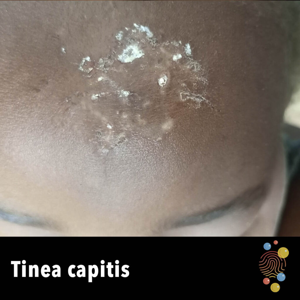
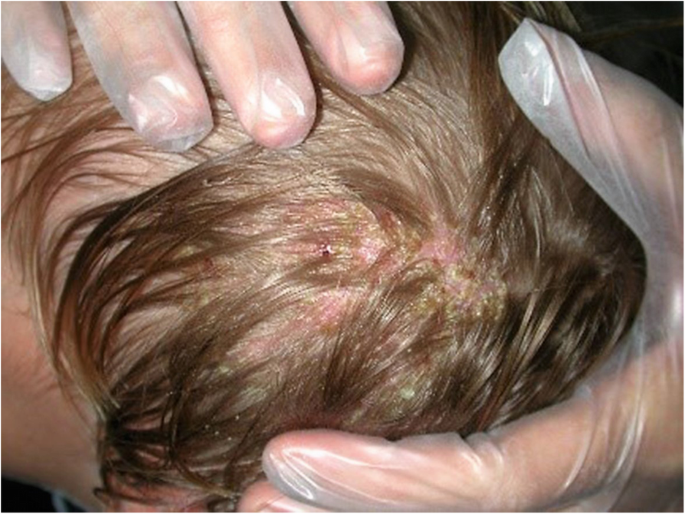
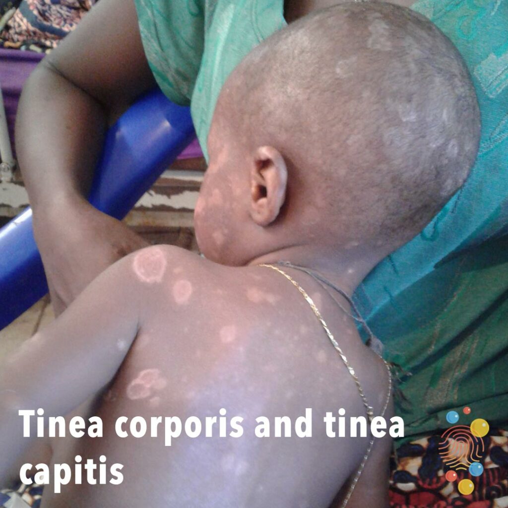
My daughter was using 200mg acyclovir pills. The bubbles continued to reappear after a few months. We tried all kinds of pills, but every effort to get rid of the virus was futile. Since then, we have moved from one hospital to another. As kerions are postulated to be an exaggerated local response to the fungus, intralesional steroids have also been proposed so as to prevent scarring and to permit possible faster regrowth of hair.My name is hoover, my 18 year old daughter, Tricia was diagnosed with herpes 3 years ago. All shared brushes, combs, and bed linens need to be thoroughly disinfected. Preventative measures of similar shampooing regimens should be carried out by close contacts as well. When infection is confirmed, all family members must be evaluated with scalp cultures regardless of clinical infection in order to reduce risk of recurrent infection without this measure.Īdjunctive therapies like 2% ketoconazole shampoo, 2.5% selenium sulfide shampoo, 4% povidone-iodine shampoo should be lathered and massaged into the scalp and left on for 5 minutes, which should be repeated 2 to 3 times per week. Newer antifungal medications like itraconazole, fluconazole, and terbinafine have been proposed as more effective given the changing face of causative organisms. Oral griseofulvin has been the gold standard of therapy since its introduction in the late 1950s. A negative culture is a more accurate indicator of disease eradication than a negative KOH stain. Confirmation of the infection can be undertaken with 20% potassium hydroxide (KOH) preparation. Tinea capitis may be suspected when a classic ringworm lesion is found elsewhere on the body. Favus is more commonly found in Eurasia and in North Africa but uncommonly in the United States and Western Europe. Favus, left untreated, can lead to scarring alopecia.
#PICTURES TINEA CAPITIS SCALP SKIN#
Tinea Capitis Ringwormįavus is a type of tinea capitis characterized by scutula, which are sulfuric-yellow concretions of hyphae and skin debris. Despite the degree of swelling, scarring is oftentimes not severe and hair regrowth is expected in most cases. Those with a cell-mediated immunity are the ones that generate a kerion, whereas those who do not have this will most likely not. This represents an immune reaction to the fungus and not a bacterial superinfection. Following 10 days from onset, the area can be severely pustulated with the rapid development of a red plaque to create a raised boggy mass. tonsurans can cause a severe inflammatory lesion known as a kerion in a localized area of the scalp.
#PICTURES TINEA CAPITIS SCALP PATCH#
The hairs get broken off a few millimeters above the skin surface and appear twisted, brittle and grayish white (gray patch type).īoth M. Ectothrix invasions are principally caused by Microsporum in which the cuticle is destroyed, which fragments into masses of spores outside of the hair shaft. Endothrix and ectothrix account for the majority of the cases. There are three recognized patterns of hair invasion: endothrix, ectothrix, and favus.


 0 kommentar(er)
